1. BONES TERMINOLOGY


    Several terms are used to refer to features and components of bones throughout the body:

    Bone featureDefinition
    articular processA projection that contacts an adjacent bone.
    articulationThe region where adjacent bones contact each other — a joint.
    canalA long, tunnel-like foramen, usually a passage for notable nerves or blood vessels.
    condyleA large, rounded articular process.
    crestA prominent ridge.
    eminenceA relatively small projection or bump.
    epicondyleA projection near to a condyle but not part of the joint.
    facetA small, flattened articular surface.
    foramenAn opening through a bone.
    fossaA broad, shallow depressed area.
    foveaA small pit on the head of a bone.
    labyrinthA cavity within a bone.
    lineA long, thin projection, often with a rough surface. Also known as a ridge.
    malleolusOne of two specific protuberances of bones in the ankle.
    meatusA short canal.
    processA relatively large projection or prominent bump.(gen.)
    ramusAn arm-like branch off the body of a bone.
    sinusA cavity within a cranial bone.
    spineA relatively long, thin projection or bump.
    sutureArticulation between cranial bones.
    trochanterOne of two specific tuberosities located on the femur.
    tubercleA projection or bump with a roughened surface, generally smaller than a tuberosity.
    tuberosityA projection or bump with a roughened surface.

    Several terms are used to refer to specific features of long bones:

    Bone featureDefinition
    diaphysisThe long, relatively straight main body of a long bone; region of primary ossification. Also known as the shaft.
    epiphysisThe end regions of a long bone; regions of secondary ossification.
    epiphyseal plateAlso known as the growth plate or physis. In a long bone it is a thin disc of hyaline cartilage that is positioned transversely between the epiphysis and metaphysis. In the long bones of humans, the epiphyseal plate disappears by twenty years of age.
    headThe proximal articular end of the bone.
    metaphysisThe region of a long bone lying between the epiphysis and diaphysis.
    neckThe region of bone between the head and the shaft.


    Continue reading »
  2. HEART ATTACK


    What is a heart attack?

    A heart attack (also known as a myocardial infarction) is the death of heart muscle from the sudden blockage of a coronary artery by a blood clot. Coronary arteries are blood vessels that supply the heart muscle with blood and oxygen. Blockage of a coronary artery deprives the heart muscle of blood and oxygen,causing injury to the heart muscle. Injury to the heart muscle causes chest pain and chest pressure sensation. If blood flow is not restored to the heart muscle within 20 to 40 minutes, irreversible death of the heart muscle will begin to occur. Muscle continues to die for six to eight hours at which time the heart attack usually is "complete." The dead heart muscle is eventually replaced by scar tissue.

    Approximately one million Americans suffer a heart attack each year. Four hundred thousand of them die as a result of their heart attack.


    Continue reading »
  3. SURGICAL SAFETY CHECKLIST

    CLICK PICTURE FOR BETTER RESOLUTION

    Example from ER Season 15 Episode 19 of the need for following a checklist to ensure consistency of measurement.


    This is a demonstration of how the WHO Safe Surgery Saves Lives Checkist functions in an operation, in this case a hernia repair. For more information on the Checklist and the Safe Surgery Saves Lives Campaign, visit www.safesurg.org.
    Continue reading »
  4. CARDIAC ARRHYTMIA


    Example of ventricular fibrillation, a deadly cardiac arrhythmia if not treated within minutes


    Simulation and electrocardiogram corresponding to the heart's normal sinus rhythm. The atrial contraction is initiated by the depolarization wave (shown in yellow), which originates from the sinoatial node and corresponds to the P wave on the ECG. After a delay passing through the atrioventricular node, the activation passes to the ventricles and produces a contraction. The QRS complex indicates ventricular depolarization (shown in yellow), while the T wave corresponds to ventricular repolarization.
    Continue reading »
  5. NASAL CANNULA (NC)


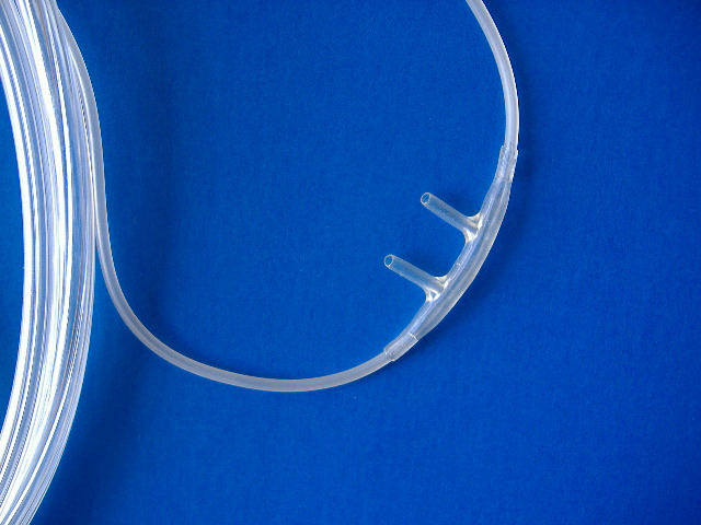
    The nasal cannula (NC) is a device used to deliver supplemental oxygen or airflow to a patient or person in need of respiratory help. This device consists of a plastic tube which fits behind the ears, and a set of two prongs which are placed in the nostrils. Oxygen flows from these prongs.[1] The nasal cannula is connected to an oxygen tank, a portable oxygen generator, or a wall connection in a hospital via a flowmeter. The nasal cannula carries 1–6 litres of oxygen per minute. There are also infant or neonatal nasal cannulas which carry less than one litre per minute; these also have smaller prongs. The oxygen fraction provided to the patient ranges roughly from 24% to 35%, or the cannula may merely supply humidified air.

    The nasal cannula was invented by Wilfred Jones and patented in 1949 by his employer, BOC.


    Supplemental oxygen

    A nasal cannula is generally used wherever small amounts of supplemental oxygen are required, without rigid control of respiration, such as in oxygen therapy. Most cannulas can only provide oxygen at low flow rates—up to 6 litres per minute (L/min)—delivering an oxygen concentration of 28–44%. Rates above 6 L/min can result in discomfort to the patient, drying of the nasal passages, and possibly nose bleeds (epistaxis).

    The nasal cannula is often used in elderly patients or patients who can benefit from oxygen therapy but do not require it to self respirate. These patients do not need oxygen to the degree of wearing a non-rebreather mask. It is especially useful in those patients where vasoconstriction could negatively impact their condition, such as those suffering from strokes. It may also be used by pilots and passengers in small, unpressurized aircraft that do not exceed certain altitudes. The cannula provides extra oxygen to compensate for the lower oxygen content available for breathing at the low ambient air pressures of high altitude, preventing hypoxia. Special aviation cannula systems are manufactured for this purpose.

    [edit]Nasal high flow

    High flows of an air/oxygen blend can be administered via a nasal cannula to accurately deliver high concentrations of oxygen.[2] Respiratory gas humidification allows the high flows to be delivered comfortably via the cannula.[3] Nasal high flow can be used as an alternative to face mask oxygen and allows the patient to continue to talk, eat and drink while receiving the therapy.[4]


    References

    1. ^ Image of nasal cannula from Glasgow Rescue Squad
    2. ^ Sim, DA; Dean, P; Kinsella, J; Black, R; Carter, R; Hughes, M (September 2008). "Performance of oxygen delivery devices when the breathing pattern of respiratory failure is simulated.". Anaesthesia 62 (9): 938–40. PMID 18540928.
    3. ^ Turnbull, B (2008). "High-flow humidified oxygen therapy used to alleviate respiratory distress". British Journal of Nursing (Mark Allen) 17 (19): 1226–30. PMID 18974691.
    4. ^ Tiruvoipati, Ravindranath; Lewis, David; Haji, Kavi; Botha, John (September 2009). Journal of Critical Care (World Federation of Societies of Intensive and Critical Care Medicine). doi:10.1016/j.jcrc.2009.06.050. PMID 19781896

    Continue reading »